14 Nov 2017

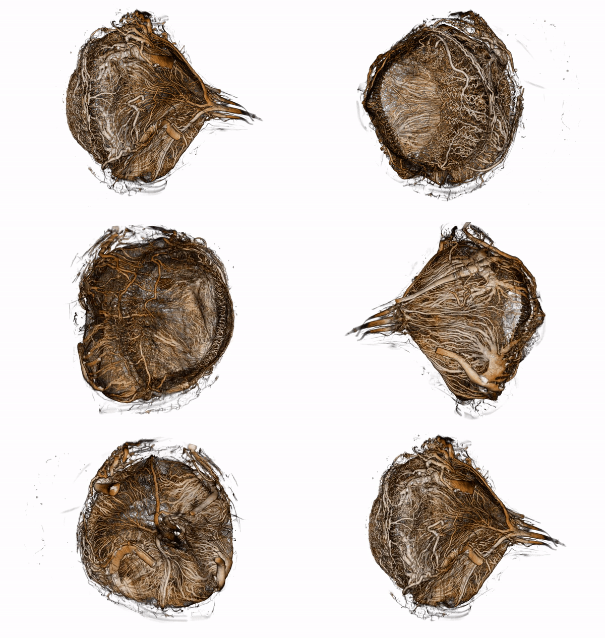
The movie shows a 3D visualization of a tomographic scan of the eye of a 5 week old Wistar rat.
The eye was instilled with µAngiofil and scanned on a SkyScan 1272 with a source voltage of 60 kV, a source current of 166 µA with camera and geometry settings leading to an isotropic voxel size of 1.25 µm.
The diameter of the eye is approximately 5 mm.
The data set was visualized in 3D Slicer, where we exported the rotation as a set of single images.
This set of images was then converted to a movie with
cat *.png | avconv -f image2pipe -framerate 24 -i - -c:v h264 -b:v 5000k -preset veryslow -pix_fmt yuv420p -vf scale=-2:1080 output.mp4
The output.mp4 movie was then converted to an auto-playing gif file with this small bash script.
01 Nov 2017

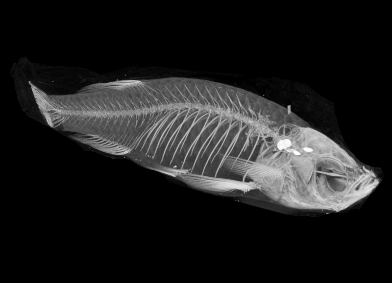
A 3D visualization of a tomographic scan of a zebrafish in native state (9 months old).
The zebrafish was scanned with an isotropic voxel size of 10 µm.
The total length of the zebrafish embryo is approximately 3 cm.
The data set was visualized using ClearVolume in Fiji.
20 Oct 2017

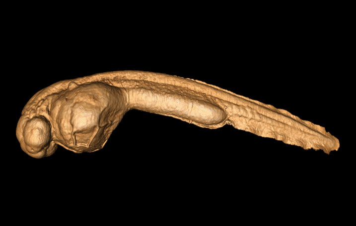
A 3D visualization of a tomographic scan of a stained zebrafish embryo (44h old).
The embryo was scanned on a SkyScan 1272 with a source voltage of 60 kV, a source current of 166 µA with camera and geometry settings leading to an isotropic voxel size of 3 µm.
The total length of the zebrafish embryo is 2.7 mm.
The visualization was made with 3D Slicer.
18 Oct 2017

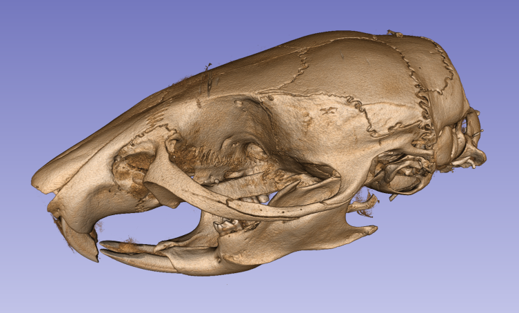
The image shows a 3D visualization of a tomographic scan of a skull of a mouse approximately five months old.
The visualization was made with 3D Slicer.
The mouse head was scanned on a SkyScan 1272 with a source voltage of 90 kV, a source current of 111 µA with camera and geometry settings leading to an isotropic voxel size of 11.25 µm.
The whole head is approximately 27 mm long.
28 Aug 2017
We scanned a fixed (4% PFA) and stained (1% Lugol solution) zebrafish.
The fish was scanned on a SkyScan 1272 at 2.5 um pixel size.


